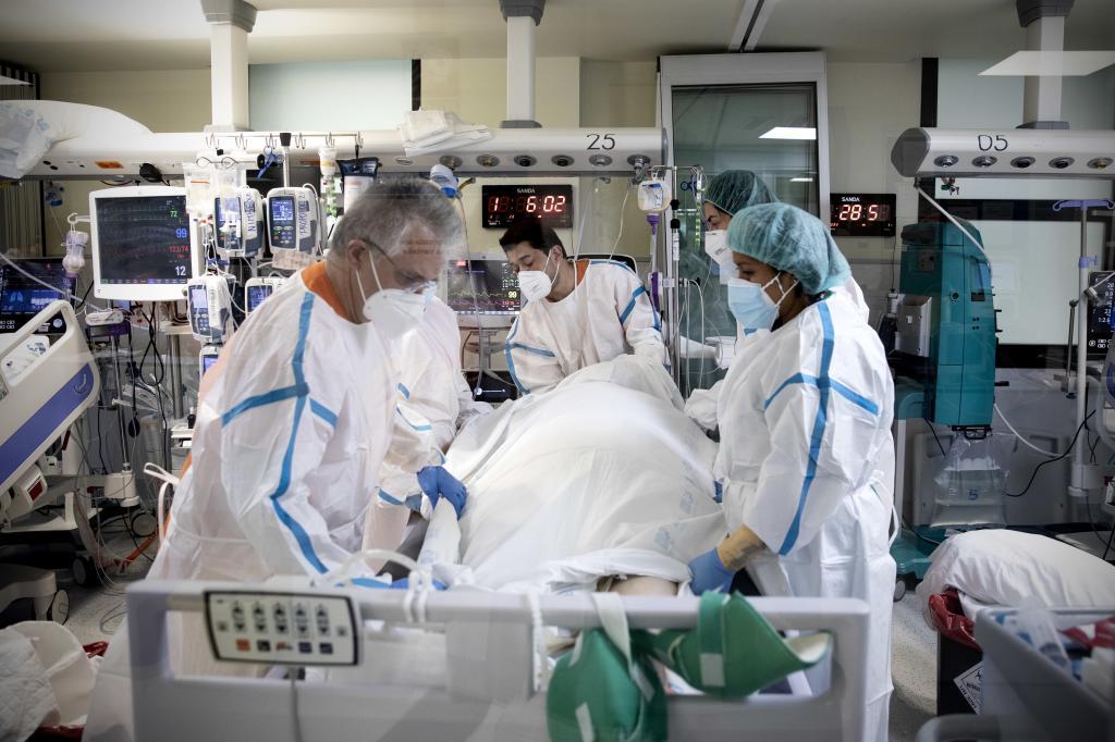
From the early days of the pandemic, the ability of Covid-19 to ravage the lungs and end the lives of many infected individuals became evident. However, until now, we did not have the details of that destruction; a map that has been developed by an international team of researchers led by Manel Esteller, a scientist from the Sant Pau Research Institute in Barcelona, whose findings are published in the journal Nature Communications.
According to Esteller, their work provides the most detailed cellular mapping to date on the deadly effects of Covid on the human lung. This definition has been achieved through the use of spatial transcriptomic techniques that make it possible to identify, while maintaining the tissue architecture, which genes are turned on or off throughout the process and at each point in the lung.
The scientist emphasizes that this is the first study that allows drawing a map of the cellular arrangement and genetic activity while preserving the lung tissue in its natural state. In previous research, it was common to have to fragment the tissue for analysis. However, in this case, the scientists were able to observe which genes were activated at a precise location in the lung, preserving the actual tissue architecture as left by the disease. "It is, in a way, like replacing a blurry aerial photograph of a city with a detailed satellite map, street by street, showing exactly which neighborhoods thrive and which are crumbling," the researchers state in a press release.
The scientists studied lung samples from 19 patients who died from Covid and compared them with healthy control lungs. This analysis not only revealed, step by step, how the infectious disease can transform healthy lung tissue into scar tissue but also uncovered therapeutic options that could be useful in halting this destruction.
The first thing the map shows is the progressive collapse of the oxygen exchange system that Covid causes in the lungs in severe cases. Thus, the cells that allow oxygen to pass from the air to the blood gradually die as the disease progresses.
Additionally, the mapping also shows the progressive transformation of healthy lung tissue into scar tissue.
"There is an increase in fibrosis around the bronchioles, which hinders respiratory movements" and gradually colonizes the tissue, explains Esteller, who also highlights that during the process, some key immune cells, such as NK cells, a first line of defense against viral infections, decrease, reinforcing the hypothesis that a failure in the deployment of these cells contributes to the lethality of the disease.
As these 'good' cells were lost, the researcher points out, the team also noted an increase in myeloid cells, related to chronic inflammation.
The scientists have also identified a molecular pathway that drives fibrosis progression.
In these cases of progressive destruction, the lung tries to heal itself, but often fails, a process that the study has also dissected.
"In these patients who did not survive Covid, we see how the progenitor or mother cells (AT2 cells) that should regenerate or create functional alveolar cells, responsible for oxygen exchange, were left halfway and could not generate the same cells," says Esteller, who recalls that some previous research by his team had already elucidated why this happened in some individuals and not in others. "The epigenetic and genetic profiles of each individual give a certain propensity to better or worse repair lung damage," he clarifies.
Much of the identified damage patterns are specific to Covid, the researcher points out. By comparing their data with lung samples from cases of pneumonia unrelated to Covid and idiopathic pulmonary fibrosis, the team demonstrated that the destruction footprint observed constitutes a distinctive signature of Covid and "not a common feature of any severe lung injury," they emphasize.
Furthermore, the study has also identified several drugs that could be useful in treating the active disease and preventing its progression. "Among them are fibrosis inhibitors targeting TGF-B1: inflammasome drugs (NLRP3 blockers); and inhibitors of the more 'belligerent' myeloid cells in the excessive immune response (such as MYB blockers)," Esteller points out.
Understanding the molecular pathways and cell types involved in Covid-associated lung destruction allows us to be better prepared for future pandemics, Esteller predicts.
Because although the study reflects "that the cellular and molecular alterations observed in people who died from Covid are different from other pneumonias or fibrosis cases," it also shows "that there are common factors."
"The use of high-resolution techniques such as the spatial transcriptomics used here allows dissecting the tissue factors altered by the virus's arrival and opens the door to more specific therapies. Additionally, viruses with similar structures trigger similar pathological cascades, therefore the study lays the groundwork for considering how the lesions induced by a similar viral agent, associated with a disease that we could call Covid-20, might be one day," he concludes.